Every year, millions of Americans notice something on their skin and wonder: Should I be worried about this? A new spot, a mole that seems different from how it used to be, a patch that will not quite heal. Sometimes the answer is nothing, but sometimes it is the earliest warning sign of the most common cancer in the United States.
Skin cancer affects more Americans than all other cancers combined. The good news is that when detected early, most forms of skin cancer are highly treatable. The challenge is knowing what to look for and knowing when a spot on your skin warrants a call to a board-certified dermatologist.
This guide walks through exactly what skin cancer can look like across its most common forms, the tools dermatologists use to evaluate suspicious lesions, and the clearest signals that it is time to make an appointment. Whether you are doing a self-exam for the first time or preparing questions for an upcoming visit, this article gives you the information you need to take your skin health seriously.
The ABCDE Rule: Your At-Home Framework for Evaluating Moles
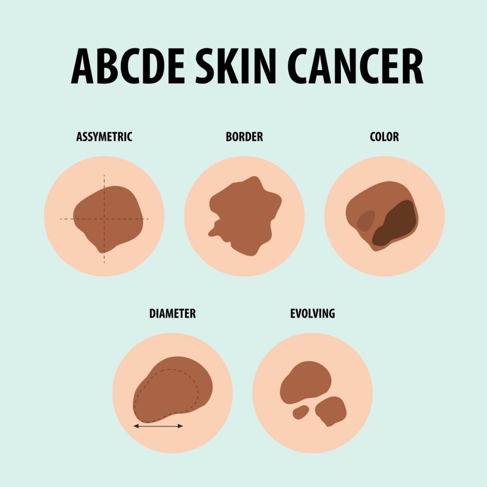 The ABCDE rule is the most widely used self-assessment tool for identifying moles that may be at risk of melanoma, the deadliest form of skin cancer. It gives you five clear criteria to evaluate any spot on your skin:
A — Asymmetry
A normal mole is typically round or oval, and both halves look the same. If you drew a line through the middle of a mole and the two halves do not match, that asymmetry is worth noting. Melanoma lesions are often irregular in shape, and no two halves will look alike.
B — Border
Benign moles have smooth, well-defined borders. A concerning mole may have edges that are ragged, notched, scalloped, or poorly defined, where the pigment seems to bleed or fade into the surrounding skin. Irregular borders are a hallmark of several types of skin cancer, particularly melanoma.
C — Color
Healthy moles are generally a single, uniform shade of brown or tan. Multiple shades within the same lesion — mixtures of brown, black, red, white, or blue — are a significant warning sign. Uneven pigmentation within a single spot should always be evaluated by a dermatologist.
D — Diameter
Melanomas are often larger than 6 millimeters in diameter — roughly the size of a pencil eraser — when diagnosed. However, they can be smaller when detected early. Any spot that seems to be growing warrants evaluation regardless of its current size.
E — Evolving
This may be the most important criterion. Any mole or skin lesion that is changing in size, shape, color, or any other characteristic — or that develops new symptoms such as bleeding, itching, or crusting, should be seen by a dermatologist. Change is the key signal your skin sends when something is wrong.
| Clinical note from Loudoun Dermatology
The ABCDE rule is a helpful starting point, but it was designed primarily to identify melanoma. Other common and dangerous skin cancers — basal cell carcinoma and squamous cell carcinoma — often look quite different and can be missed by self-exam alone. This is why an annual full-body skin check with a board-certified dermatologist remains the gold standard for skin cancer detection. |
The Three Main Types of Skin Cancer and What Each One Looks Like
Skin cancer is not a single disease. There are three primary types, each with distinct visual characteristics and levels of risk. Understanding the differences can help you identify warning signs that the ABCDE rule alone may not capture.
Basal Cell Carcinoma (BCC)
Basal cell carcinoma is the most common form of skin cancer, accounting for roughly 80 percent of all cases. It typically grows slowly and rarely spreads to other parts of the body — but if left untreated, it can invade surrounding tissue, nerves, and bone.
BCC most often appears on sun-exposed areas: the face, scalp, ears, neck, and hands. It can take several forms:
- A pearly or waxy bump, often skin-colored or slightly pink, that may have visible blood vessels on the surface
- A flat, flesh-colored or brown scar-like lesion
- A pink growth with raised edges and a crusted center that may bleed easily
- A sore that repeatedly heals and then reopens
Many patients first describe BCC as a “pimple that will not go away” or a spot that bleeds when they wash their face. If you have had a non-healing sore for more than a few weeks, it should be evaluated.
Squamous Cell Carcinoma (SCC)
Squamous cell carcinoma is the second most common skin cancer. It carries a higher risk than BCC because it can spread to lymph nodes and internal organs if left untreated. People with weakened immune systems, those who take immunosuppressant medications, and people with a history of organ transplants are at elevated risk.
SCC often develops from actinic keratoses — rough, scaly, pre-cancerous patches caused by years of sun exposure. It commonly appears on the face, ears, lips, back of the hands, and lower legs, and can look like:
- A firm, red nodule or rough, scaly patch that does not heal
- A new sore or raised area on an old scar
- A rough, wart-like growth that may crust or bleed
- A flat lesion with a scaly, crusted surface
Melanoma
Melanoma is the least common but most dangerous form of skin cancer. It develops in the melanocytes — the cells that give skin its pigment — and has a much higher risk of spreading to other organs if not caught early. Early detection is critical: the five-year survival rate for melanoma detected at its earliest stage is over 98 percent. That rate drops dramatically once the cancer has spread.
Melanoma can arise from an existing mole or appear as a new, unusual growth. It can develop anywhere on the body, including areas rarely exposed to the sun such as the soles of the feet, the palms, under fingernails, and even on the eyes. Signs include:
- A large brownish spot with darker speckles
- A mole that changes in color, size, or feel, or that bleeds
- A small lesion with an irregular border and portions that appear red, pink, white, blue, or blue-black
- A dark streak under a fingernail or toenail
- A lesion that itches, burns, or is tender
| Important: Skin cancer is not always dark-colored
A common misconception is that skin cancer always appears as a dark or black spot. Basal cell carcinoma, one of the most common forms, is often skin-colored, pink, or translucent. Amelanotic melanoma — a particularly dangerous variant — lacks pigment entirely and may appear as a pale, pink, or red patch. Never assume a light-colored growth is safe simply because it is not dark. |
How Often Should I Get a Full-Body Skin Check?
 One of the most common questions dermatologists hear is: “How often do I really need a skin exam?” The answer depends on your personal risk factors, but general guidelines provide a clear starting point.
The baseline recommendation
The American Academy of Dermatology recommends that adults perform monthly self-examinations and have a full-body skin check by a board-certified dermatologist at least once a year. Annual exams are especially important because many areas of the body, such as the back, scalp, behind the ears and between the toes, are difficult or impossible to see properly on your own.
Higher-risk patients should come more frequently
Your dermatologist may recommend more frequent visits if you have one or more of the following risk factors:
- A personal history of skin cancer, including basal cell or squamous cell carcinoma
- A family history of melanoma
- Many moles, or unusual moles (known as atypical or dysplastic nevi)
- A history of severe or repeated sunburns, especially in childhood
- Fair skin, light hair, or blue or green eyes
- A history of significant sun exposure or tanning bed use
- An immunocompromised status due to disease or medication
- A history of organ transplant
Patients in these categories are sometimes seen every three to six months. Your dermatologist will help establish a monitoring schedule tailored to your skin and history.
Do not wait for your annual exam if something changes
The annual exam is a baseline, not a reason to delay. If you notice any new or changing spots, a sore that will not heal, or anything that concerns you between appointments, call your dermatologist. Skin cancer can progress quickly, and early intervention almost always leads to better outcomes.
What Happens During a Skin Cancer Screening Appointment?
 Many people avoid or postpone skin checks simply because they do not know what to expect. A full-body skin examination is quick, non-invasive, and can be genuinely life-saving. Here is exactly what happens during a screening at Loudoun Dermatology Associates.
Before the exam
You will be asked to remove your clothing and change into a paper or cloth gown. It helps to arrive with clean, dry skin — no heavy lotions, nail polish, or makeup on areas you want evaluated. Bring a list of any spots or areas you have noticed or have questions about.
During the exam
A board-certified dermatologist or physician assistant will systematically examine your skin from head to toe, including the scalp, between the toes, the soles of the feet, and other areas you cannot easily see yourself. The exam typically takes 10 to 20 minutes.
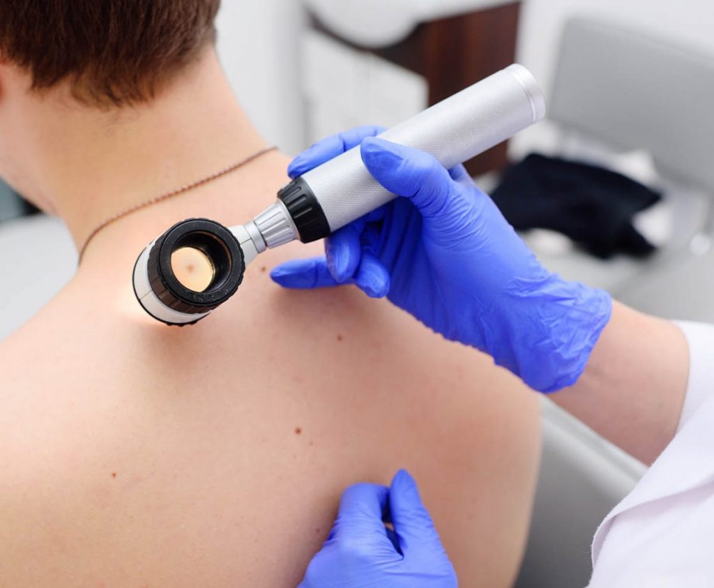
Your provider may use a dermatoscope — a handheld magnifying device with a built-in light — to examine individual lesions more closely. Dermoscopy allows dermatologists to see structures beneath the skin surface that are invisible to the naked eye, significantly improving diagnostic accuracy.
After the exam
Most patients leave with either a clean bill of health or a recommendation to monitor specific spots at a future visit. If your dermatologist finds something suspicious, they will discuss the findings with you and may recommend:
- A watchful waiting approach with a follow-up appointment in a few months
- A biopsy to obtain a tissue sample for laboratory analysis
- Immediate treatment if a lesion appears highly concerning
A skin biopsy is a minor in-office procedure that involves numbing the area with a local anesthetic and removing a small tissue sample. Results typically return within one to two weeks.
| A note on skin checks at Loudoun Dermatology
Loudoun Dermatology Associates has been providing skin cancer screenings to patients in Loudoun County since 1997. Dr. Van Ha and our team of certified physician assistants have examined over 30,000 patients and have the expertise to evaluate all skin types, tones, and ages. Annual skin checks are covered by most insurance plans. |
Can Skin Cancer Appear in Places Not Exposed to the Sun?
 This surprises many patients: yes, skin cancer — including melanoma — can and does develop on parts of the body that receive little or no sun exposure. While ultraviolet radiation from the sun is the leading cause of most skin cancers, it is not the only cause.
Locations to check beyond the obvious
Most people are aware they should watch for suspicious spots on their face, arms, and chest. But skin cancer can also develop in:
- The soles of the feet and between the toes
- The palms of the hands
- Under or around the fingernails and toenails (subungual melanoma)
- The genitals and buttocks
- The scalp, especially under the hair
- Inside the mouth and nasal passages (mucosal melanoma)
- Inside the eye (ocular melanoma)
Melanomas that develop in non-sun-exposed areas can be especially dangerous because they are often discovered later, when they are more advanced. Subungual melanoma — appearing as a dark streak under a nail — is frequently mistaken for a bruise or fungal infection.
UV exposure is not required for melanoma
While sun damage drives the majority of skin cancers, some melanomas are caused by genetic mutations unrelated to UV radiation. This is why people with darker skin tones, who have more natural protection against UV damage, are still at risk for skin cancer — particularly in non-sun-exposed locations. Acral lentiginous melanoma, the type most common in people of Asian, African, and Hispanic descent, typically develops on the palms, soles, and nail beds.
Regardless of your skin tone or sun exposure history, a full-body skin check with a dermatologist is the most thorough way to catch skin cancer early, wherever it might appear.
What Does a Biopsy Involve and How Do I Prepare?
If your dermatologist recommends a biopsy, it is natural to feel some anxiety. Understanding exactly what the procedure involves can make a significant difference in how you feel going in.
Why a biopsy is recommended
A skin biopsy is the definitive way to determine whether a lesion is cancerous, pre-cancerous, or benign. While an experienced dermatologist can often make a visual assessment with strong confidence, a biopsy provides a cellular-level analysis that removes all uncertainty. It is a routine procedure performed thousands of times each week in dermatology offices across the country.
Types of skin biopsy
The type of biopsy your dermatologist recommends depends on the size, location, and suspected nature of the lesion:
- Shave biopsy: A blade is used to shave off the surface layers of the growth. Used for raised or superficial lesions. Results in minimal scarring.
- Punch biopsy: A circular tool removes a small, cylindrical core of skin that includes deeper layers. Typically requires one or two stitches. Used to assess lesions that may extend deeper into the skin.
- Excisional biopsy: The entire lesion is surgically removed along with a small margin of surrounding normal skin. Recommended when melanoma is suspected or the lesion is large.
What to expect on the day of your biopsy
The procedure takes place in the office and usually lasts less than 15 minutes. Your dermatologist will:
- Clean and numb the area with a local anesthetic injection — this is typically the only discomfort involved
- Remove the tissue sample using the appropriate method
- Place a bandage over the site
- Provide aftercare instructions for keeping the wound clean and protected
Most patients return to normal activities immediately after. The biopsy site may be slightly tender for a few days.
Understanding your biopsy results
Results typically return from the laboratory within one to two weeks. Your dermatologist will contact you to review the findings and, if needed, discuss next steps. A benign result means no further treatment is required for that lesion. A malignant or pre-malignant result will be followed by a discussion of treatment options, which may include additional excision, topical medications, or referral for more advanced care.
| What happens if skin cancer is confirmed?
Most skin cancers detected early are treated with an outpatient surgical procedure at Loudoun Dermatology Associates. Basal cell and squamous cell carcinomas are often removed by excision or Mohs surgery, which removes cancerous cells layer by layer while preserving as much healthy tissue as possible. Early-stage melanoma is typically treated with wider excision. Your dermatologist will walk you through every option and recommend the approach best suited to your diagnosis. |
When to Make an Appointment: A Clear Decision Framework
If you are unsure whether your skin concern warrants a dermatology visit, use this framework:
Book an appointment if:
- You have a mole or spot that meets any of the ABCDE criteria (asymmetry, irregular border, multiple colors, diameter over 6mm, or evolution)
- You have a sore, wound, or spot that has not healed within four to six weeks
- A lesion bleeds without being injured, or bleeds repeatedly
- You have not had a full-body skin check within the past 12 months
- You have a personal or family history of skin cancer
- You have fair skin and a history of significant sun exposure or tanning bed use
- You have a new spot that looks different from all your other spots — the “ugly duckling” sign
Seek same-week attention if:
- A lesion is growing rapidly
- A previously stable mole suddenly bleeds, itches, or becomes painful
- You notice a dark streak under a fingernail or toenail that has not resulted from injury
When in doubt, make the call. A spot that turns out to be benign costs you 20 minutes of your time. A spot that turns out to be early-stage skin cancer, caught at that appointment, could save your life.
About Loudoun Dermatology Associates
Loudoun Dermatology Associates has served the Leesburg, Virginia community and all of Loudoun County since 1997. Led by Dr. Van Ha, a board-certified dermatologist who completed his residency at Johns Hopkins Hospital, our practice offers comprehensive annual skin cancer screenings, diagnostic biopsies, and both general and surgical dermatologic care for patients of all ages, skin types, and ethnicities.
If you have a mole, spot, or skin concern you would like evaluated, we welcome you to schedule an appointment. Early detection is the most powerful tool we have, and we are here to use it with you.
This article is intended for educational purposes and does not constitute medical advice. Consult a board-certified dermatologist to evaluate any skin concern.